© Ula Krzanowska / The Hoof Architect 2024–2026. All rights reserved.
Unauthorized reproduction, redistribution, adaptation, or derivative works (including redrawn diagrams or similar concepts without attribution) prohibited without written permission.
This content, including original observations, biomechanical interpretations, and diagrams, was first publicly presented by the author at the 4th International Congress on Equine Podiatry, Jerez de la Frontera, Spain, May 16–18, 2024.
Introduction to Part 4: The Hoof is Not a Monolith
In the previous parts of this series, we covered macro-level dorsopalmar distortions and the limb biomechanics responsible for them. However, to truly understand these patterns, we must look deeper.
The hoof capsule is not a uniform block; it is a complex assembly of tissues with different mechanical properties that distort at different rates.
In this part we are going to cover the details of the internal anatomy associated with the specific distortion patterns and focus on specific areas of potential pathology due to overload.
Hoof Distortions on the Micro Level
Hoof capsule distortions are not random defects of the horn. They represent mechanical adaptations of a complex biological structure to the loading pattern generated by the limb.
By micro-level distortions we refer to specific changes occurring within the individual structures that build the hoof capsule.
These micro-level distortions organize into patterns that eventually manifest as macro-level distortions affecting the entire functional unit — both the horn capsule and the internal structures.
Hoof shape is an expression of load distribution and capsule deformation, not a result of a few geometric parameters.
Depending on the biomechanics of the distal limb, the hoof may distort in different ways.
Several important principles should be considered:
- The hoof capsule is not uniform, and it matters which structures are loaded and how they are loaded.
- Different components of the hoof possess different mechanical properties and therefore distort at different rates and in different ways.
- Some elements collapse, others remodel, and some shift position and change shape.
Among the structures that undergo particularly prominent and relatively rapid changes in shape — apart from the coriae — are:
- the digital cushion
- the coronary cushion
- the collateral cartilages
These structures are closely interconnected with the horn capsule. When the horn capsule distorts, these tissues distort as well, and later influence the position and angle at which horn is produced.
Different areas of the hoof distort due to different mechanical factors.
Coronary Groove and Wall Formation
Distortions of the coronary band on a micro level were partially discussed in Part 3 of this series.
The shape of the coronary groove is influenced by the way the hoof is loaded and has a direct effect on the thickness and quality of the wall it produces. In other words, the coronary groove reflects the interaction between the internal structures and the horn capsule during load.
This interaction influences the overall hoof morphology and may alter the way the entire hoof is loaded, creating a feedback loop that can become a vicious cycle.
The more vertical the coronary groove becomes, the less cushioning effect it provides and the thinner the wall it produces. Thin walls are less rigid and therefore more susceptible to infection, mechanical damage, and semi-permanent deformation.
The coronary groove tends to distort differently in different regions of the hoof. These distortions correlate directly with load distribution and macro-level distortion patterns.
The greater the upward shearing force between the bone and the wall, the more vertical and narrow the coronary band becomes. This phenomenon affects both the dorsal area and the area around the quarters, extending approximately to the point where the palmar processes of the coffin bone end.
Behind this point, the coronary band tends to experience less shearing and instead moves together with the soft tissues that form the caudal hoof structures.
As explained in Part 3, distortions resulting from laminitic damage can further exacerbate coronary band distortions. Many chronic laminitic feet exhibit very thick dorsal walls while the toe quarters remain thin and weak.
Wall thickness is influenced by many factors, with genetics playing an important role. However, in many cases variations in wall thickness appear to be strongly related to biomechanics.
Examples below demonstrate how significantly wall thickness may vary depending on the shape of the coronary groove.
Coronary Cushion and Digital Cushion
The coronary band is mechanically connected with the structures that form the caudal part of the hoof. Consequently, when the digital cushion displaces, the coronary band displaces with it, and vice versa.
In feet that are considered ideal, a common pattern can be observed: the coronary groove tends to become gradually narrower as it progresses from the dorsal wall — where it is thickest — toward the heels.
When load distribution within the hoof becomes disrupted, the width and height of the coronary groove may change as a result of vertical shearing forces between the wall and the bone.
The location of this overload depends heavily on limb conformation and often correlates with the DCA value.
Feet characterized by high DDFT tension and a narrow DCA typically show the greatest stretching of the coronary band in the dorsal area. In contrast, hooves with upright pasterns and wide DCA values often display compression in the central region of the coronary band.
Based on these observations, this may represent one of the aspects of central hoof overload.
The point at which the coronary groove appears to be most vertically stretched roughly corresponds to the end of the palmar processes of P3. Externally, this region often coincides with the area where growth rings display signs of wall compression.
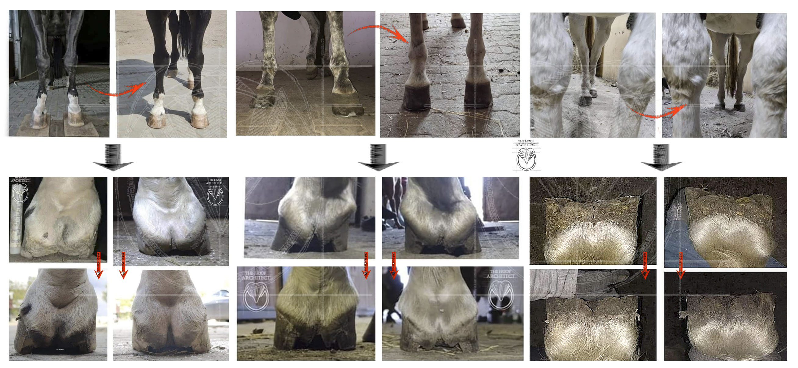
Two limbs with completely different biomechanics – the first one was the upright fetlock case (the horse had some SDFT injury) and on the right a limb with a tendency for a dropped fetlock and more flexion in the coffin joint.
Behind the point where the palmar processes end, the bone no longer provides the same structural influence. The coronary groove therefore becomes more relaxed and tends to move upward together with the collateral cartilages instead of significantly stretching.
Research by Paige Poss has demonstrated that the heel buttress is tightly connected with the collateral cartilage without the coronary cushion positioned between them.
In many cases this area corresponds with the location where quarter cracks develop. This appears to be the point where the coronary band is pushed upward the most relative to the collateral cartilage, which may be one of the factors predisposing the hoof to quarter cracks, as described in the teachings of Hans Castelijns.
Quarter Cracks and Load Distribution
Based on these observations, quarter cracks appear to be commonly associated with central or caudal overload of the hoof.
Central overload often corresponds with wide DCA values.
When quarter cracks occur unilaterally, they tend to develop on the side that is shunted upward, which typically has a wider DCA than the opposite side.
When they occur bilaterally, they are often found in hooves where both sides display wide DCA values.
This pattern appears to correlate with:
- upright fetlock conformation
- DDFT laxity
The situation may be further exacerbated by downward displacement of the digital cushion, which is often associated with open-heel shoes.
Such shoeing may increase pressure under the wings of P3 while simultaneously reducing caudal hoof expansion during load. This even further increases compression between the collateral cartilages and the coronary band.
Comparison of Wide and Narrow DCA Hooves – Internal Anatomy
A deeper examination reveals consistent anatomical patterns that occur in hooves with wide and narrow DCA values.
In the example shown above:
The upper hoof represents a narrow DCA hoof, associated with DDFT contracture and hypo-extension (chronic flexion) of the coffin joint.
The lower hoof represents a wide DCA hoof associated with an upright pastern, broken back alignment and a greater range of extension in the DIP joint.
Several differences can be noted:
- different thickness and shape of the coronary groove in the areas marked with arrows
- differences in sole shape
- different frog apex morphology — round and deep in narrow DCA hooves versus elongated and flat in wide DCA hooves
Additional differences include:
- the relationship between the internal heel and the dorsal wall
- underrun in narrow DCA
- more parallel in wide DCA
- heel bulbs
- pulled downward and forward in narrow DCA hooves, producing an arch-shaped, concave bottom
- pushed upward and stretched backward in wide DCA hooves, producing a flatter or even convex bottom
Frog width may be influenced by several factors, but certain tendencies are often observed:
- in narrow DCA hooves, the frog base tends to be wider with straighter edges
- in wide DCA hooves, the frog tends to become stretched, elongated, and narrower, often displaying a characteristic change in shape beneath the navicular bone area.
Another significant difference is observed in the collateral grooves:
- in narrow DCA hooves they tend to remain relatively straight
- in wide DCA hooves they often collapse beneath the navicular bone area
This feature may be particularly important both as an indicator of internal balance and as a potential source of clinical problems.
A comparison of a normal (left), wide DCA (middle) and narrow DCA (right) foot internal anatomy.
Caudal Hoof Elongation
Caudal structures are attached to relatively rigid components of the horn capsule such as the bars, heels, and frog. Because of that, they distort together.
The characteristic elongation of the caudal part of the hoof — which can sometimes create the impression of heel contraction — can often be observed in wide DCA hooves.
In many cases such hooves are not necessarily too narrow. Instead, the caudal structures may simply be excessively elongated, although both conditions may also occur simultaneously.
The digital cushion has limited volume. When it becomes elongated, it also becomes narrower.
It is often suggested that the digital cushion can regenerate and increase in volume. While some degree of regeneration may occur, observations frequently indicate that displacement and change of shape are more prominent processes than true tissue regeneration.
When the digital cushion becomes elongated, it may become mechanically weaker and less effective in its functional role.
Collateral Grooves and the Navicular Region
One area that has drawn particular attention is the region corresponding with the characteristic indentation observed in the collateral grooves, especially in wide DCA hooves.
Whilst this indentation may be a normal occurence to certain degree, its more pronounced shape along with specific bar shape appears to correlate with several common clinical problems.
The shape of the collateral grooves varies significantly between wide and narrow DCA hooves.
This indentation may be associated with forces exerted by P2 onto the navicular bone.
The feature may appear:
- in both collateral grooves
- in only one groove
- or with varying degrees of prominence
In cases involving mediolateral deformities, the indentation may be present on one side and absent or less pronounced on the other. Its presence often correlates with the DCA value on each side of the hoof.
Sections Through the Collateral Groove Region
Sections along the deepest part of the collateral grooves of narrow (top) and wide (bottom) DCA feet.
In hooves with high DDFT tension (narrow DCA):
- the digital cushion beneath the navicular bone tends to be thicker
- it is positioned more forward and downward
In wide DCA hooves:
- the digital cushion beneath the navicular bone is often flattened and compressed
- it is stretched backward and upward
These findings appear to repeat consistently depending on DCA value and limb conformation, although individual variation is always present.
In wide DCA hooves, bruising beneath the navicular region is relatively common. In such cases the digital cushion may be extremely thin and the bars of the horn capsule may press directly into the DDFT.
This region is also where Paige Poss identified the branch of the digital palmar nerve branch.
Compression in this area may therefore result not only from direct pressure but also from the stretching of the entire bottom of the horn capsule.
In mechanical terms, the heels tend to pull backward while the toe pulls forward. The navicular region lies between these opposing forces and may function as a mechanical fulcrum, concentrating stress.
I suspect that compression of this area is not only related with direct pressure but also from the stretching of the whole bottom of the horn capsule. I believe this creates an extra tensile strain with the navicular area in the middle acting as a fulcrum pressing down, which would be the cause of the persistent ‘end of bar’/’bar-sole junction’ cracks. These cracks can be painful and cause lameness (considering the location of the nerve this is not a surprise), may bleed and get infected. This is a very common location of bruises and hoof abscesses. In my experience, unloading and floating the whole heel and bar in a ‘reverse arch’ configuration (to relieve that stretching effect) has instantly and significantly improved comfort and led to those cracks growing down over time in several horses.
Chronic and recurrent bar cracks related to central hoof capsule overload. They can cause lameness and abscesses.
Contraction of the heels makes that problem even more complex, when descending pastern collapses the heel buttresses, pressing the bars forward into the navicular region:
Bar crack and folded compressed bar related to the central area overload and compression between the navicular bone and the horn capsule combined with narrow heels and pastern descending onto the heel and bar. In this case it was possible to restore better heels geometry and solve the problem.
Area of central overload related to the compression of the navicular area.
In my little personal cadaver limb study (more details in another article), filling the whole bottom of a wide DCA foot with 35 shore A DIM and putting a pressure of about 300 kg has created a significant pressure spot on the pressure mat and later the dissection has shown a significant bruising in that specific area, which also corresponded with some DDFT pathology localised in this area.
A comparison of the collateral groove - navicular area relationship. Left: narrow DCA hoof with DDFT contracture - the caudal hoof suspended by the tendon. Right: wide DCA hoof of and upright fetlock limb. Direct compression of the structures between the navicular bone and horn capsule.
The narrow DCA hoof with DDFT contracture presented in the illustrations above is also a disfunctional one, with the area of compression and pathology localised at the dorsal/cranial area. This is related to the excessive DDFT tension shifting the weight onto the toe, preventing the extension of the DIPJ and disrupting breakover mechanics. The common pathologies relates to this aspects are stretch of the dorsal coronary groove, thin dorsal wall, dorsal wall cracks, flare, white line problems around the toe area, thin and flat sole, P3 remodeling and abscesses.
A study of the wide DCA, upright fetlock limb with fetlock varus. An obvious are of pathology localised under the lateral navicular region, clear signs of chronic compression in the area of the digital palmar nerve branch location. This is a very common abscess area, especially common in this type of feet. Changes in the DDFT are probably chronic. Bruises are most likely post-mortem, created during the hydraulic press test after filling the bottom of the foot flash with 25 shore A DIM. The corresponding pressure mat reading attached. There were no high spots on the sole.
Common hoof related oversimplifications
Some widely repeated explanations attempt to attribute hoof distortions primarily to a small number of geometric parameters. However, long-term observations of hoof morphology suggest that these relationships are far more complex.
What about the internal arch spoken about so often?
This type of distortion is not uncommon (especially in mediolateral distortions!). I believe trimming this type of feet with unloading the quarters may exacerbate the problems (this was my experience too) and they need to be treated in a manner focused on diminishing that stretching of the bottom horn capsule aspect.
Another important implication of the distortion within the collateral grooves is that assessing P3 orientation based on the depth of the collateral grooves is likely to be inaccurate. The further the DCA from the normal range, the more likely the error of external assessment markers.
What about upright pasterns being considered favourable by some schools?
Whilst some horses with true upright conformation and DDFT tension being in balance with the fetlock position may indeed serve as a good biomechanical example, attempting to artificially increase fetlock orientation in any given horse may create problems of many natures.
One of the patterns I often observe is that horses that have undergone certain injuries or have certain orthopedic problems (tendon or suspensory ligament injury, dorsal fetlock joint arthritis, deep thrush for example) tend to adopt a more upright fetlock position as a compensatory posture without any corresponding change in hoof angulation. These horses often present numerous chronic and very difficult to resolve hoof problems related to the aspects described above.
Apart from that, anegdotic evidence provided by experienced orthopedic vets and riders suggest, that this type of horses tend to perform well only until a point, with a high tendency to break suddenly.
What about the influence of PA on the shape of solar arch and collateral grooves?
If PA was indeed the primary determinant of load distribution within the hoof, we would expect to observe consistent distortion patterns corresponding to its value. In fact, studying thousands of feet over many years, I have not found this to be the case. Hooves with similar PA values may present very different solar arch shapes and collateral groove patterns.
Increasing or decreasing PA is likely to shift the weight in more or less predictable way but it must be remembered that this shift is relative and a starting point is individual for each limb, not global!
Moreover, in many cases achieving and maintaining specific PA values may be difficult or impossible due to the same conformational factors that determine the shape of the hoof capsule. In this sense, PA may often represent an expression of limb conformation rather than a primary cause of hoof distortion, which indicates that correlation may sometimes be mistaken for causation when interpreting certain hoof problems.
My long-term observations of hoof morphology changes in live horses show how much more complex this topic is.
DCA and x-rays: uncommon aspects to look at
Except for the DCA measurement, we can assess some of the features corresponding with internal foot distortion on x-rays.
On top what would be considered ideal, normal DCA value.
Straight collateral grooves with the peak around the area of the coffin bone wings. (Of course depending on how long the palmar processes are it may vary.)
On the left top: a total caudal collapse. Almost no depth to the collateral grooves. Wide DCA.
Below left: a collapse of the central area and partially of the caudal too. Wide DCA
On the right top: overloaded toe, caudal hoof suspended due to high DDFT tension, no compression of the horn capsule below the navicular area, narrow DCA
Right bottom: compression of the navicular area, collateral grooves extending above the wings of P3. Wide DCA.
Collateral grooves can be seen on x-rays. Depending on how clean the hoof is and whether it has been trimmed, whether thrush is present it can be more or less accurate - I suggest that trimming the frog overlapping the collateral grooves first and marking the bottom with barium paste may make the assessment more precise.
In wide DCA feet, there a pronounced indentation in the bars is present, the bottom of the collateral groove is often located above/behind the P3 wings. It seems like the caudal horn capsule is whole shunted up against P3. P3 wings and navicular bone are embedded deep within the horn capsule.
In narrow DCA feet the collateral grooves tend to be more smooth and rounded, without dips and with the deepest area closer to the center of the foot.
A set of examples of different scenarios depending on the limb conformation and the relationship between the caudal structures, coronary cushion and collateral grooves have been marked.
Top: narrow DCA; middle: wide DC and central overload; bottom: wide DCA and central/caudal overload.
Bone Remodeling: The Long-Term Record
The coffin bone (P3) is the last structure to change. While soft tissues shift in seconds, horn capsule can distort in days/weeks, bone takes years to remodel. I believe the distortions we see in the coffin bone—such as bone loss and remodeling or changes in the palmar processes—are highly related to the quality of the SADP (Suspensory Apparatus of the Distal Phalanx) and long-term postural habits, rather than just the orientation of the hoof against the ground.
Coffin bones also respond to this load distribution.
A set of x-rays showing similar P3 distortion patterns related to similar conformations of the limb. This topic requires much more research.
Conclusion
The observations described above illustrate how hoof distortions often emerge at a very small structural scale before they become visible in the overall shape of the hoof capsule. Changes in the collateral grooves, coronary band, solar arch and within the caudal hoof appear to reflect the internal loading patterns of the foot rather than simple geometric parameters such as hoof angle or palmar angle alone.
Studying large numbers of feet over time shows that the hoof is not a rigid structure defined by fixed angles. Instead, it is a continuously adapting capsule responding to the forces generated by limb conformation, movement patterns and compensatory postures. Small distortions accumulate gradually as the tissues adapt to these forces, and the visible morphology of the hoof often represents the external footprint of those internal mechanics.
Because of this, interpreting hoof form through a limited set of measurements or idealized geometric values may lead to misleading conclusions. Similar values of commonly used parameters can be associated with very different internal distortion patterns within the hoof capsule.
Observing the distribution and character of these distortions in the whole hoof capsule may therefore provide important clues about how the limb is actually loading the foot.
Understanding these micro-level changes is an important step towards a more realistic biomechanical interpretation of hoof morphology.
In this context, parameters such as DCA may be interpreted less as static measurements of hoof form and more as morphological records of the loading patterns acting within the limb.
In Part 5 we will finally put all of this together and look at the actual 3×3 matrix of dorsopalmar hoof types based on fetlock position and DDFT tension.
Special thanks to Paige Poss for all the support, long hoof-geek discussions and for our collaborative dissections with many lightbulb moments.


Comments
Post a Comment