Demistifying equine limb deformities and how they refer to hoof capsule distortions - introduction

Let’s get one thing straight: every horse’s leg is crooked to some degree.
Symmetry does not exist in nature. Straight horses do not exist. No leg is straight, no bone is straight and no joint is straight. Sometimes crookedness is obvious even for an untrained eye and sometimes it’s hard to spot.
It is not possible to illustrate and describe all possible variations of conformational and postural deviations (or rather it would require a whole book to present them all). Instead, my goal is to set a common language for talking about equine crookedness, so everybody can understand them and won’t need to learn by heart anymore.
Theoretically, horses’ legs can be deformed in almost infinite amount of variations (according to Daisy Bicking it is over 700 possibilities1). Practically, those deformations seem to appear in certain specific combinations and patterns, with rather rare exceptions. Anyway, there is so much more to it than just toed-in, toed-out, bow legged or knock-kneed!
In this article I’ll try to make this complex topic a bit clearer without oversimplifying it - as oversimplifying is actually what I assume to be the cause of all this chaos and misunderstanding.
This is an introduction and theoretical preparation to the more practical part which is described in part II, where I am describing 4 types of hoof deformations using the terminology described in this part. Understanding those 4 types of deformations and their causes gives a lot of answers of what problems you may be dealing with and how and when to correct them.

Conformation or posture?
First of all, we need to distinguish between conformational and postural faults. Usually both occur simultaneously and are often confused for one another. Because what do we mean by ‘toed-in’? Is it about the inward rotation of the whole limb (posture), an inward rotation of the cannon bone or pastern bone (rotational deformity), or maybe fetlock varus (angular deformity)? Or maybe a combination of the above?
Why does it matter?
Probably this kind of oversimplification is the reason why the common patterns of hoof deformities are not so clearly identified. With this approach, it would seem as if the conformation does not correlate with hoof capsule distortions that much, as each of the examples above (toed-in) would present with a completely different shape of a hoof capsule. Each would also need a different farriery approach and allow different possibilities for correction.
We need to understand that what we see when looking at the horse from in front is a result of its conformation (angular and rotational deformities) AND posture.
Conformation can and often does affect the horse’s posture. It affects the way the hoof is loaded and therefore the way it grows, which affects the way the horse is adjusting its posture to be comfortable with the given hoof balance. (Fig. 24.)
In adult horses, posture does not affect the conformation, but it does influence the load distribution over the hoof capsule and can lead to bone remodeling over time.
In foals and growing horses, posture does affect conformation as it impacts the way the bones fuse.
Conformation is something we are not able to change in a fully developed horse. Some of the deformations in the bones and joints can be corrected at a very young age.
Posture, on the other hand, may change at every stage of a horse's life, depending on its comfort and development and there is much more we, as hoof care professionals (and other equine specialists), can do about it.
Every leg can be deformed or posturally adjusted independently.
The way the hoof is loaded, the way it grows and the way it deforms is a result of the horse's conformation AND posture. Conformational and postural deviations can add up to each other or be balancing each other out, creating more or less deformed hoof capsule.
Some types of deformities have a bigger impact on hoof distortions than the others, regardless of their severity.
Conformation should be assessed at the axis of the limb, whilst posture - at the axis of the whole horse - more on that later.
CONFORMATION
Generally speaking, conformational crookedness consists of angular and rotational deformities in the limb.
Every leg, every bone and every joint can be deformed independently. Many of those deformities are congenital not corrected on time but they may also appear during growth or even later, as a result of injury.
ANGULAR DEFORMITIES
Angular deformities can occur in the joints (valgus/varus type of deformities: when the angles between joint surfaces and long axis of the bones below and above the joint don’t match each other) or in the bone itself (‘banana’ shape: when long axis of the bone is bent or offset: when one joint surface is shifted mediolaterally in reference to the other).
.jpeg) Fig. 3. Front view:
Fig. 3. Front view:- Straight front limb
- Carpus valgus: medial deviation of carpal joint
- Carpus varus: lateral deviation of carpal joint
- Fetlock varus: lateral deviation of fetlock joint
- ‘Banana’ shaped pastern often interpreted as fetlock valgus
- Offset knee
.jpeg) Fig. 4. Rear view:
Fig. 4. Rear view:- Straight hind limb
- Tarsus valgus: medial deviation of tarsal joint
- Tarsus varus: lateral deviation of tarsal joint
- Fetlock varus: lateral deviation of fetlock joint
- ‘Banana’ shaped pastern often interpreted as fetlock valgus
Images illustrating only angular deformities, not the whole horse posture. Angular deformities must be assessed in the limb axis. Note that rarely ever a horse would be standing with both limbs facing perfectly forward.
Offset joint*
There are some cases where offset in the joint is not a result of an asymmetrical shape of the bones but rather a subluxation in the joint itself.

Fig. 5. X-rays of right front limbs of two different horses:
- Medial offset in the coffin joint - the coffin bone is shifted medially in reference to the short pastern
- Lateral offset in the coffin joint - the coffin bone is shifted in reference to the short pastern X-rays taken by Ada Majocha DVM.
Sometimes the offset in the joint can be dependent on hoof balance and can be corrected to some point.
Joint tilt*
Joint tilt does not equal to angular deformity - when a horse is standing, the leg may look straight and the deformity will be apparent only when the leg is being bent. In such case the long axis of both bones may be aligned but the joint surface will not be parallel to this axis. It will be apparent in movement, though, and affect the swing phase of the leg (paddling, dishing)
.jpeg)
Fig. 6. Left front limb, fetlock joint, front view.
- Straight joint with no tilt.
- Straight joint with a lateral tilt: this will be apparent when the leg bends.
- Angular deformity in the joint: fetlock varus.
- Angular deformity in the bone: ‘banana’ shaped long pastern bone.
- Angular deformity in the joint: fetlock valgus. Very uncommon type of deformity, often confused with no. 4.
The difference between 1&2 and 4&5 will be apparent when the leg bends.
Angular deviation means that the angles between bones’ axis and the joint surface don’t match.
.jpeg)
Fig. 7. Paddling and dishing* due to medial and lateral tilt in the carpal joint. This can apply to any joint but the longer the bones creating it, the more pronounced the deviation.
*Paddling and dishing can be also caused by inward or outward rotation of the whole limb. Here we speak of a limb facing straight forward and still exhibiting a swing phase deviation.

Fig. 8. 4 left front cannon bones. Most cannon bones present a lateral slope in their distal epiphysis (clear in the bones 3 and 4). If the pastern bone is straight, such combination will result in fetlock varus - like in the picture to the right.
Bones to the left are the property of doctor Ric Redden, bones to the right belong to Rood and Riddle Equine Hospital in Lexington.
*This term seems a bit misunderstood to me, as I have seen it being used for describing multiple different phenomena: sometimes as joints’ position in reference to the ground. However, this is dependent on hoof balance, horses’ posture and fetlocks’ position and I don’t think this parameter should be mentioned as a conformational factor.
ROTATIONAL DEFORMITIES
Rotational (spiral) deformities occur in the bones, when one epiphysis is twisted in reference to the other. Doctor Ric Redden calls them intorsion (when distal epiphysis is twisted medially in reference to the proximal) or extortion (when distal epiphysis is twisted laterally in reference to the proximal)
.jpeg)
Fig. 9. Set of long pastern bones used for the study. On the left: ‘banana’ shaped bone with almost no twist. In the center: Pastern bone without significant angular deviation but with severe medial twist (intorsion). On the right: Pastern bone with a pronounced ‘banana’ shape and significant intorsion.
Bones belong to doctor Ric Redden and were photographed at his clinic.
.jpeg)
Fig. 10. Front view:
- Straight front limb
- Medial twist in the pastern
- Medial twist in the cannon bone
- Lateral twist in the radius
- Lateral twist in the radius combined with medial twist in the cannon bone. This will be apparent in movement and will likely force the horse to land laterally first, as well as in no. 4.
Images illustrating only rotational deformities, not the whole horse posture and without angular deformities. Rotational deformities must be assessed in the limb axis. Note that rarely ever a horse would be standing with both limbs facing perfectly forward.
.jpeg)
Fig. 11. Rear view:
- Straight hind limb
- Medial twist in the pastern
- Medial twist in the cannon bone
- Lateral twist in the tibia
- Lateral twist in the tibia combined with medial twist in the cannon bone
Images illustrating only rotational deformities, not the whole horse posture and without angular deformities. Rotational deformities must be assessed in the limb axis. Note that rarely ever a horse would be standing with both limbs facing perfectly forward
Rotational and angular deformities occur simultaneously.
Theory vs practice
In theory, every joint can be straight, have a tilt or angular deformity both ways. Also in theory, every bone can be straight, twisted both ways, bent or offset.
In practice however, I suspect that there are some ranges and directions in which specific bones can deform, which would be related with the forces playing part when a horse is standing and moving.
*With that in mind, still deformities in longer bones or in joints created by longer bones (cannon bone, radius, long pastern) will be much more pronounced than those including short bones (short pastern, coffin bone). Thus it is usually spoken of carpus and fetlock varus/valgus and I've never heard of pastern joint or coffin joint varus/valgus, even though they most likely occur.*
For example, according to doctor Ric Redden, fetlock joints would only naturally deform as varus angular deformity, and the very few that present as true valgus would be caused by human intervention.
There are some configurations of deformities that may be optically perceived as fetlock valgus, though.
Banana type deformity of the pastern as well as lateral coffin joint offset, especially combined with base wide posture, can create an impression of fetlock valgus. True fetlock valgus, though, would cause paddling of the digit in the swing phase, as it would mean that joint space in the fetlock would be tilted to the inside. A vast majority of horses present a lateral tilt in fetlock joints, no matter if the limb is straight or if it has fetlock varus or ‘banana’ shaped pastern.
Significant outward twist of the whole limbs (no matter if it's just due to posture or outward rotational deformity in the radius) will give an impression of fetlock valgus, even if in fact there is fetlock varus! The same goes the other way round - if the whole limb is twisted to the inside, it will look like fetlock varus. It is because the fetlock joint will be assessed at an angle and not at its axis. That's why it's crucial to judge the deformities in the leg axis, not the whole horse axis.

Fig. 12. Illustration showing the same (straight) front limb viewed at different angles. The fetlock joint, because of the way it’s built, may create an impression of angular deformity when viewed at an angle.

Fig. 13. Illustration showing the same (straight) hind limb viewed at different angles. The fetlock joint as well as the hock, because of the way they are built, may create an impression of angular deformity when viewed at an angle.
.jpeg)
.jpeg)
The second one looks like severe fetlock varus, but this is caused by the cannon bone intorsion. His fetlock is also almost straight, with a slight banana shape in the pastern and mild inward rotation of the hoof capsule.
Both horses have quite symmetrical hoof capsules.
The same applies to offsets and rotational deformities in the bones.
For example, I have never encountered a carpal joint offset medially.
Cannon bones and pastern bones mostly present as straight or twisted medially (especially in pastern bones this spiral may be really pronounced). I have never seen a pastern bone with extorsion, but the research is ongoing. Radius bones seem to often have lateral twist.
It seems it is not coincidental that certain angular deformities go hand in hand with certain types of twists. The most severe intorsion in the pastern bones occurs in horses with either lateral ‘banana’ shaped pasterns or lateral coffin joint offsets. Fetlock varus usually combines with straight and symmetrical pastern bones.
*I have a theory that it has to do with the leverage occurring especially when a foal is doing tight turns - the inside leg is subjected to much bigger twisting forces than the outside leg, that is why those forces don’t balance each other out.

Fig. 15. A horse with significant intorsion of the pasterns. Note how the turning affects the position of the cannon bone in reference to the hoof capsule: the twisting force is visible with naked eye.
The more lateral leverage (banana or offset in the lower limb), the more twisting forces. With fetlock varus, the leverage is much shorter, so those cases are not subjected to such twisting forces and don’t have the tendency to develop this twist.
Is there a correlation between keeping young foals in small paddocks and the occurrence of pastern intorsion?*
Together with Ada Majocha we spoke to multiple top level farriers and podiatrists to find out what their observations and experiences were. We found out that opinions differed a lot and that there was not a one consistent approach to this topic.
According to my current knowledge, this topic has not been researched comprehensively yet.
POSTURE
Posture can be neutral (all cannon bones perpendicular to the ground - what about the carpal/tarsal valgus/varus cases, though?) or compensatory. It may be independent to conformation: a horse with almost perfectly straight limbs can stand in a compensatory way and a severely crooked-legged horse can stand neutral. Each limb can be adjusted independently.
Regarding mediolateral balance, limbs can be twisted in or out, spread wide or placed narrow. It is usually talked about neutral, base wide or base narrow posture (to be assessed on a standing horse from in front as a relationship between the position of the center of the hoof's base of support and the center of the axis of the elbow or stifle joint - my suggestion of how to assess, to be verified) but it’s not precise enough. Base wide posture can be a result of a horse just spreading its legs wide but can also be caused by an outward rotation of the whole limbs.
Both situations will have different effects on the distribution of load over the hoof capsule, mainly because they affect the direction of the fetlock descend. This factor is so important that it deserves a whole separate chapter.
.jpeg)
Fig. 16. Front limbs, front view. All drawings show straight limbs with the only variable being the changing posture.
.jpeg)
Fig. 17. Hind limbs, rear view. All drawings show straight limbs with the only variable being the changing posture.
CONFORMATION + POSTURE
What we ultimately see in a horse is a sum of angular and rotational deformities and posture.
We should add components from all 3 graphics to get the end result.

.jpeg)
These graphics show only some of the possible angular and rotational deformities that occur in horses. Imagine the amount of all possible combinations. With all this understanding now, what does ‘toed-in’ mean?
As said before, conformational and postural components add up to each other, exacerbating or partially canceling each other out.
The ultimate factors that determine whether and in what way the hoof capsule is going to be deformed are base width and direction of fetlock descend, both of which are a resultant of conformation AND posture.
Angular and rotational deformities between the fetlock joint and the coffin bone have the biggest impact on hoof distortions, regardless of their severity (severe intorsion in the cannon bone will create less deviations in load distribution over the hoof capsule, than milder intorsion in the pastern).
FETLOCK JOINT
When it comes to hoof capsule shape and deformities, I find fetlock joint to be the most important factor.
This joint is really amazing!
It is so unique, like no other joint in the equine body.
It is sometimes said that the fetlock is what makes a horse a horse2.
If I were to tell one factor that decides about ‘hoof conformation’, the fetlock would be my bet.
The fetlock is a hinge joint allowing minimal rotation, adduction and abduction but with the highest range of motion (flexion and extension) of any equine joint (up to 120°). It absorbs concussion, stores energy and stabilizes the distal limb2.
It is built as a suspension bridge that is unable to support the load until the tendons and ligaments tense to support the cannon bone and the whole body above2. It does not lock in a fixed position but instead is suspended in a sling made of deep and superficial flexor tendons, suspensory ligament and other ligaments.
Its neutral position is dependent on the tension over DDFT and SDFT, which partially depends on hoof balance but also on flexor muscles tension. It also depends on the amount of load the limb is bearing.
*That is why it is pointless to assess the ratio of limb lengths by looking at the height of the carpus joints! If one leg is bearing more weight, the fetlock will drop lower and the carpus join will follow.*
Descending fetlock is shifting the weight of the equine body from the bones to the tendons and ligaments. Tensing up DDFT gives more suspension to the caudal part of the coffin bone (pulling its palmar processes up) but at the same time descending pastern is a huge lever compressing the caudal part of the hoof. This is, what I assume, what creates change in DCA value, which is basically about how deep the caudal part of the coffin bone is embedded in the hoof capsule.

Fig. 20. Descending fetlock changes the pressure distribution over the hoof capsule. On one hand the lower the fetlock, the more compression to the caudal structures of the hoof. On the other hand, the longer lever arm caused by descending fetlock, the more tendon (which means also DDFT) tension, giving suspension to the caudal part of the coffin bone. This, I believe, is what makes the DCA become narrower over time in hooves with dropped fetlocks. On the other hand, fetlock descending less than physiological (like for example with SDFT contracture), puts less tension on the DDFT (less suspension at the area of the palmar processes of the coffin bone) along with less compression at the caudal area of the hoof. This creates hooves with low PA, high heels and narrow back of the foot (migrated, wide DCA hooves - link).
Fetlock joint and hoof capsule deformities
Angular and rotational deviations between the fetlock and the coffin bone are the most significant when it comes to occurrence of severe hoof capsule deformities.
If a horse has a twist in the pastern, has angular deformities in the fetlock or coffin joint or is placing its limbs abnormally wide or narrow, it will result in a fetlock not descending inline with the hoof capsule, but medially or laterally.
If the fetlock is descending inline with the hoof capsule, it fits in between the collateral cartilages and descends into the digital cushion and frog stay. However, if it descends towards one of the sides, it encounters one of the collateral cartilages and has to push it out to descend. This in time semi-permanently changes the position of the cartilages, soft tissues building the caudal hoof, coronary band, direction of the wall growth and whole hoof capsule morphology. Unaddressed, it can lead to sheared heels, quarter cracks and collateral ligaments injuries as well as coffin bone remodeling.

Fig. 21. Hoof capsule with coffin bone aligned with the pastern axis and fetlock descending symmetrically between the collateral cartilages. Red color in the picture to the right shows the area compressed by the descending fetlock.

Fig. 22. Fetlock descending symmetrically between the collateral cartilages vs fetlock descending offaxis. Red color shows where the pressure goes when the fetlock descends.
Hoof capsule deformities
Hoof capsule deformities can be temporary (they disappear once the force creating them is removed), semi-permanent (they can be reversed in time) and permanent (irreversible).
Hoof capsule is a viscoelastic structure.
When the fetlock descends to one of the sides, the hoof capsule deforms and gets back into almost the same shape as before once the force is removed (temporary deformation). When this action is repeated continuously over a longer period of time, the hoof is slowly exhibiting creep - a continued deformation of a viscoelastic material after the load has reached a constant state. This is how sheared heels, flares and asymmetry in the hoof capsule occurs (semi-permanent deformation).
Those changes are usually at least partially reversible over time, provided that we are able to change the forces acting on the hoof capsule.
If, however, uneven load and blood circulation persists for a long period of time, coffin bone can get remodeled or the collateral cartilage ossified and those changes will be permanent and irreversible - but that does not mean we are not able to address the capsular deformities at all.
*Uneven growth is not yet a deformation
When the hoof is loaded unevenly, it creates uneven blood flow, which leads to uneven growth. If one whole side of the hoof just grows faster, it only needs to be trimmed level to get back into balance.

Fig. 23. X-ray and venogram of the same hoof. Note the difference in angulation of the side walls (the steeper side is experiencing more load|) and how that corresponds with the length of the solar papillae (they are shorter on the overloaded side due to compression). The load distribution affects the blood flow and blood flow affects the growth rate. That is why an unevenly loaded hoof grows unevenly (the less steep side will grow faster.)
X-rays and venograms performed by Doctor Redden at his farm.
When the disbalance is not addressed for a long time, it can lead to capsular deformities, when some parts of the hoof shift positions in reference to the other ones. In those cases it needs a very conscious approach and a lot of understanding of why and how those semi-permanent changes occur to (sometimes only partially) reverse them - or rather to enable parts of the hoof to get back into their places over time.
Those semi-permanent deformations include deformations of the walls, shifts in the coronary band shape and position - which presents as sheared heels, asymmetry in side walls angles and thickness, flares and curling under. If addressed properly, load distribution over the hoof can change, leading to coronary band position change, leading to change in direction and thickness of growing walls.*

3 directions of deformations
As a result of mediolateral imbalance, hoof capsule can deform in 3 directions (usually all 3 occur simultaneously at different ratios):
Horizontal bend

Vertical (transversal) shift

Sagittal twist

Sheared heels - is it all about the fetlock descend direction?
Some people believe sheared heels have to do with the way the horse is landing, which I completely disagree with. It is sometimes stated that sheared heels occur when one collateral cartilage gets pushed up in reference to the other one, which I partially disagree with.
Collateral cartilages are firmly attached to the coffin bone and it’s not really possible to push one of them up independently on the coffin bone position - only the caudal part can be deformed independently to the rest. In most hooves with sheared heels it’s not only the caudal hoof deformation, the shearing actually starts around the toe quarter area and extends up to the heel bulbs. What is interesting, in those hooves the coffin bones are tilted in the opposite direction to the way the heels are shunted: the palmar process on the side of the ‘pushed-up’ heel bulb is lower than the opposite one. This means it is sunk deeper in the hoof capsule, which strongly correlates with DCA.
DCA on the pushed-up side is higher than on the opposite side (high DCA hooves actually present features of a hoof that would have 2 pushed-up heels!)
Fig. 28. The same hoof from lateral and medial view with mildly sheared heels. Note the difference in DCA values on both sides: side that is being pushed up has higher DCA value, with the coffin bone embedded deeper into the hoof capsule on this side.
This means that one palmar process of the coffin bone is embedded deeper in the hoof capsule than the other. On the other side it’s sometimes even like the hoof capsule was pulled down in reference to the side of the coffin bone.
Is it due to the asymmetrically descending pastern pushing the adjacent wing of the coffin bone down? Or maybe due to less DDFT tension on that side as the tendon has to travel a shorter distance to this side of the coffin bone than it has to the other one? I hope to find the precise answers in time.
Anyway, we can (and in most times should) address this issue.
‘Sheared heels’ is actually a twist between the cranial and caudal part of the hoof capsule

That is also why a flat trim is not able to fix the problem. I believe that most cases of severely sheared heels are ‘man made’ - by trimming a twisted hoof capsule flat! In those cases, if you only address the disbalance in the joints (trim flat to balance the cranial part of the hoof, under the coffin bone), you exacerbate the disbalance in the heels. If you only address the disbalance in the heels (trim flat to get even length/height of the heels), you exacerbate the disbalance in the joints!
.jpeg)
Fig. 29. Two most common ways of addressing the deformed hoof balance. Neither of them is really helping correct the deformity. If so, is leaving it as it is the best way? Or maybe we should choose the way to trim based on the most severe issue we are dealing with?
Or what if we could combine both of the trims together, to get the benefits of both? We’ll focus on that in part III, which may be in the form of an article or a webinar.

Fig. 30. An example of severely sheared heels in both front feet of the same horse and how this deformation can be corrected over time.
Sheared heels issue is neither about the distance of each heel bulb from the ground, nor about their length ratio. Instead, it is about the position of the heels and the coronary band in reference to the vertical axis of the hoof capsule.

Fig. 31. A deformed hoof with sheared heels. It is neither the ratio of heel lengths, nor the distance of each heel bulb from the ground that determines that, though.
How to determine if the heels are sheared?

Fig. 32. A precise way to determine how much heels are sheared. The dashed line is a bisector of the angle between side walls directions: the axis of the hoof capsule. Thick black line is perpendicular to this axis and is a reference for measurements.
Yellow and blue dots are the points from which the last heel tubules grow.
Red arrow is parallel to the axis of the hoof capsule and is showing a difference in heel bulbs position in reference to the hoof capsule axis.
Why do we need this measurement?
Sometimes in the process of rehabilitation, when hoof capsule deformities are getting reversed, it may be difficult to judge whether heel bulbs are getting more symmetrical or not: because all parts of the hoof capsule are changing positions. This is a reliable way to verify it.

Fig. 33. An example of a left front hoof with sheared heels during the rehabilitation process. Sometimes it may be hard to judge the heel bulbs symmetry with naked eye, especially when the whole hoof capsule has shifted. Red arrows show the shift in heel bulbs position in reference to the hoof capsule axis.


Fig. 35. As mentioned before, when the fetlock descends towards one side of the hoof (not aligned with the hoof axis), it doesn’t smoothly fit between the collateral cartilages but instead it omits one cartilage and gets in the way of the other one. It pushes its axial part up, in time changing its position to more vertical permanently and making it look like the whole cartilage on this side is pushed up - whilst it’s just that it’s unfolded and positioned more vertically which makes it look higher. At the same time, the off-axis descending fetlock is shifting the CoP towards the side it descends to, leading to vertical (transversal) shift of the hoof capsule.

Fig. 36. A mildly asymmetrical left front hoof capsule with fetlock descending medially due to pastern intorsion. Note the asymmetry in the collateral cartilages and how the medial one is more vertical. The descending fetlock is pushing its upper part towards the medial side. Dissection performed by Paige Poss at her farm in Arizona.

Fig. 37. Pictures from the same dissection performed by Paige Poss. The hoof is not severely deformed, yet the asymmetry in the cartilages is clearly visible.

Fig. 38. Illustrations showing how the difference in the collateral cartillages heights occurs. It’s not really about one of them being pushed up but rather compressed by the hoofwall and descending pastern, which leads it to becoming more vertical. On the bottom photos of the same hoof: the one on the left is after appropriate corrections, the one on the right is before.
Heels can also be shunted horizontally - instead of being pushed up, one gets pushed back, or up and back. This is due to the horizontal bend of the hoof capsule and is really common in hind hooves of type III - more on that in part II.
Direction of fetlock descend and quarter cracks
In severe cases, especially with quarter wall deformation, descending fetlock will push the upper part of the cartilage out, making it push into the coronary band and creating tensile forces in the upper part of the wall, which leads to quarter cracks.
Fig. 39. Fetlock descending medially, pushing the upper part of the medial cartilage up and towards the medial side, which creates tensile forces in the medial quarter wall. Dissection performed by Paige Poss at her farm in Arizona
*Edit on 9.10.23: Can ringbone or other thickening of the pastern bones contribute to quarter cracks?
Asymmetrically descending fetlock pushes the upper part of the adjacent cartilage out and puts it in a more vertical position. I believe that this, combined with the fact that the coffin bone and collateral cartilage is embedded deeper in the hoof capsule on this side (according to Hans Castelijns DVM this is the main cause of quarter cracks) and CoP being shifted to the same side the fetlock descends, leads to vertical compression of the adjacent side wall, which also becomes more vertical and is subjected to tensile forces in it's upper part. This shift along with the compression leads to change in direction of hoof wall growth in reference to the coronary band (coronary band gets more vertical), due to which the wall gets thinner in its cross section. In my opinion there is less intertubular horn in reference to the thicker wall on the other side. This also explains why those walls are so much weaker, prone to cracks and infections and often have problems with holding up the nails.

Fig. 40. Transverse section of a hoof with sheared heels. Note the shape of the coronary band and its reference to adjacent wall thickness. The coronary band shape is dependent on the forces acting on the walls: on the more compressed (more vertical) side, the coronary band is also more vertical. This is partially the cause of the cartilage being embedded deeper into the hoofwall on this side.
This theory also makes it clear why we hear that many of those quarter crack cases heal when barefoot and tend to crack again when shod - barefoot hoof can freely deform under the pressure of the fetlock and the compressed part will sink into the soft ground. A flat steel shoe combined with a flat trim is preventing the vertical shift of the wall, so its only displacement under pressure can take place mediolaterally. If the fetlock is descending towards the medial side and the medial quarter wall is ‘trapped’ between the shoe and the pastern, it would displace medially at the coronary band and that creates significant tensile forces leading to cracks. (With a proper trim, however, those cases can be efficiently rehabilitated in steel shoes.)

Hoof deformations and landing pattern?
Unlike commonly stated, I see no correlation between the way horses are landing and the way their hooves deform. Horses with pronounced lateral landing may have either lateral or medial heel shunted up and either lateral or medial side of the hoof capsule overloaded. Changes in landing pattern do not necessarily lead to changes in hoof morphology. There are some angular limb deformities that would force the horse to land laterally first even when the medial side of the hoof capsule is too high. Correcting the imbalance will in some cases cause more pronounced lateral landing and still lead to improvement in hoof capsule morphology and horse’s soundness. We’ll dive more into the correlation between conformation//posture and landing patterns in the next part.
The study performed by Jenny Hagen (Hagen et al. 2017) has shown that there is only a weak correlation between initial hoof-ground contact and mediolateral location of the CoP during midstance.
According to my observations, the way the horse is standing and the way its limbs are loaded during the loading phase are what predominantly determines the shape of the hoof capsule.

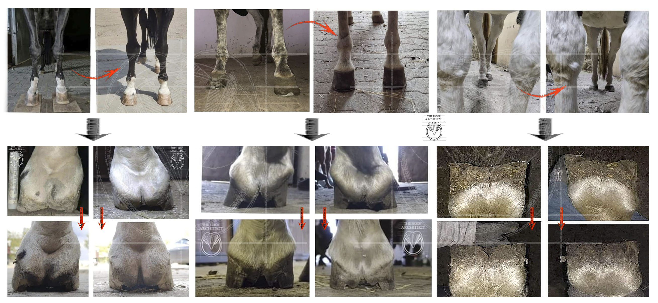
Fig. 42. A collage of examples how hoof deformation can be reversed with appropriate farriery.
Summary
Once we have a decent understanding of limb deformities, postural adaptations and mechanisms of hoof distortions and have established a common language to speak about it, we can proceed to part II - 4 crooked hoof types.
References:
Daisy Bicking, Foundation Hoofcare Handbook
Part I: Operative Orthopedics of the Fetlock Joint of the Horse: Traumatic and Developmental Diseases of the Equine Fetlock Joint Larry R. Bramlage, DVM, MS
Hagen J, Mäder D, Wuttke W, Vogel M. Immediate, short and long-term effects of hoof trimming on hoof-ground contact in the horse at the walk. Australian Equine Veterinarian 2017;36(1)
Hagen J. Mediolateral “Balance” – Scientific insights in the context of every day hoof care and shoeing Institute of Veterinary Anatomy and Horseshoeing School, Clinic for Horses, Leipzig University, Germany
—-------------
Special thanks to Ada Majocha DVM for hundreds of hours spent on discussing hooves.
Special thanks to Doc Ric Redden DVM, Paige Poss, Hans Castelijns DVM, Sammy Pittman DVM, Simon Curtis FWCF, Simon Moore FWCF, Raul Bras DVM, Daisy Bicking, the #UMF Group Members and many others for support and inspiring conversations that led me to creating this material.


Comments
Post a Comment