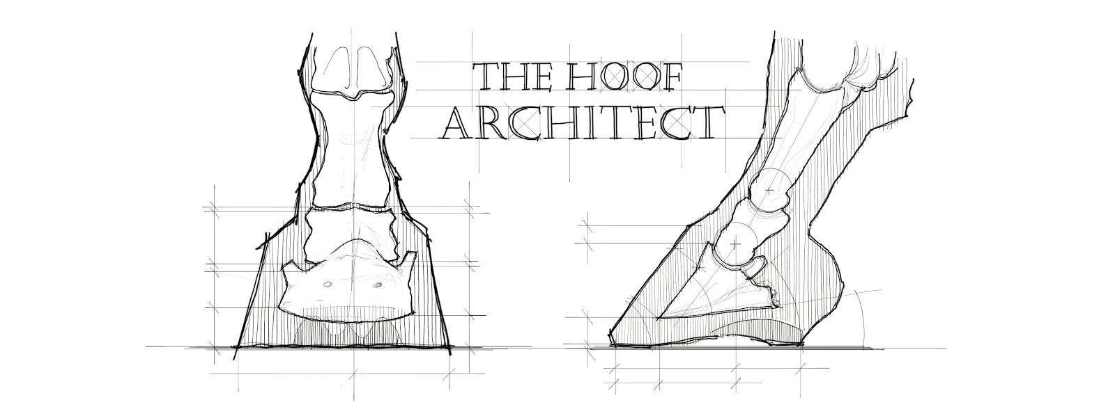
DCA and the Missing Aspects of Dorsopalmar Balance (Part 3 - Range of motion of the DIPJ and hoof morphology)
© Ula Krzanowska / The Hoof Architect 2024–2026. All rights reserved. Unauthorized reproduction, redistribution, adaptation, or derivative works (including redrawn diagrams or similar concepts without attribution) prohibited without written permission.
This content, including original observations, biomechanical interpretations, and diagrams, was first publicly presented by the author at the 4th International Congress on Equine Podiatry, Jerez de la Frontera, Spain, May 16–18, 2024.
Introduction to Part 3
In previous parts (Part 1, Part 2), the concept of DCA (Dorsal hoof wall – Coronary band Angle) was introduced as a potential indicator of internal load distribution and hoof capsule distortion patterns. Macro-level distortions were explored using simple cube models, and the role of posture, phalangeal alignment, DDFT tension and fetlock joint biomechanics was highlighted as a major influence on capsule shape.
This part will focus specifically on how the range of motion in the coffin joint (DIPJ) - its habitual alignment, extension, flexion, and their limits - directly affects coronary band and caudal structures shape and position, which in turn impacts DCA measurements and overall dorsopalmar balance.
Factors Affecting Hoof Morphology
There are numerous internal and external factors at play when it comes to determining hoof morphology:
- External:
Environment, properties of the surface the horse moves on, workload, shoeing and trimming.
- Internal:
Weight of the body relative to the hoof size, overall balance and symmetry of the body, ratio of the components building the hoof capsule (like wall thickness), genetics (bone shapes, quality of the tissues), development, nutrition and overall health (affecting horn quality and SADP quality - major factor!).
Body - hoof link
Conformation and postural adaptations seem to be major factors determining how the load is going to be distributed over the hoof capsule, which as a result determines its shape and morphology to a large degree. Their influence over the hoof is often mentioned but in many cases the talk is more general and vague.
Hoof capsule morphology appears to reflect the long-term mechanical equilibrium of the distal limb, primarily determined by the interaction between fetlock suspension, phalangeal alignment and baseline DDFT tension.
Differences in the shapes of the bones, lengths, ratios and elasticity of tendons and ligaments, muscle tension, fascia, body weight (and more) do not only affect the weight distribution in a standing horse but may as well lead to disruption in the timing and proportions of the deceleration, stabilization and acceleration components of the stance phase, altering the CoP trajectory, strain duration, load share between structures and breakover biomechanics. These ultimately determine in which areas of the hoof capsule the load is going to accumulate most heavily and which areas are going to be relatively unloaded.
In many cases locomotion biomechanics tend to correlate with the traits observed in a standing horse but they may as well differ enough to produce different outcomes for horses similarly aligned when standing.
Two of the locomotion components which can vary largely between horses and which (according to my observations) play a huge role in shaping hoof morphology - are the stance phase (maximum flexion in the DIPJ) and the breakover phase (maximum extension in the DIPJ).
DIPJ range of motion, coronary band position and caudal structures shape
The coronary band position is not fixed; it reacts to the skeletal movement above it.- Extension Mechanism:
- Flexion and Loading:
Ranges of flexion and extension in the DIPJ are determined by the conformation of the individual animal, its standing and moving posture, hoof geometry, applied weight, ground properties, etc.
 |
Coffin joint extension and flexion, and coronary band shape and position. Caudal structures and coronary groove follow P2. |
Defining abnormality is challenging, as it is difficult to specify precisely what is normal and where is its limit. However, regarding hoof distortions, my understanding would be that abnormality encompasses everything that creates further distortions. When the DIPJ extends or flexes beyond the range that was expected for it by the horn capsule, it pushes its way forward or backwards and creates that space - neither the horn capsule nor the caudal structures are likely to prevent that. If this repeats constantly, it leads to the distortion of the dorsal and/or caudal area of the hoof, displacing and distorting the coronary band and the caudal structures.
According to my observations, hooves with a wider range of extension in the coffin joint tend to have a more open DCA and the coronary band descending in front. Limited range of extension tends to correlate with DCA on the narrow side. If that range becomes abnormally limited, both loading and breakover biomechanics may change and the hoof capsule will need to adjust accordingly.
Hooves with a lot of coffin joint flexion (which depends on hoof angle and MCJ suspension) often have the coronary band pushed down in the caudal area, together with the caudal structures, whilst abnormally upright pastern is going to allow the caudal structures to migrate up and back.
I propose that the habitual range of flexion and extension in the coffin joint (DIPJ) is one of the primary determinants of coronary band deformation patterns, which subsequently influence DCA values and dorsopalmar hoof capsule morphology.
DIPJ extension range and breakover patterns
Depending on the range of DIPJ extension, the alignment of the phalanges during breakover may vary significantly, which as a result affects hoof morphology.
 |
The moment of maximum extension of the DIPJ affects the shape of the dorsal coronary band.
I have been observing that certain hooves are prone to flaring even when they have relatively short toes, some will grow very long and still maintain a straight dorsal wall and some will even develop a bullnose instead of a flare, despite a very overgrown toe and low heels:
 |
| 3 ‘long toe’ feet with completely different hoof morphology. |
Analyzing movement phases of differently conformed horses in slow motion in walk has led me to interesting observations. Breakover patterns show large diversity between individual animals, which often tends to correlate with the traits visible in their standing posture and past injuries. Excessive/insufficient DDFT tension and variations in position of the fetlock joint are the factors that may have the most influence.

Breakover biomechanics vary a lot among horses, depending on conformation, balance, and numerous other factors.
Large DIPJ extension range
A lot of flexibility in the coffin joint coupled with a longer toe lever can delay breakover by creating more hyperextension in the coffin joint before the breakover starts.If the range of coffin joint extension is wider than ‘normal’ the more acute angle between P2 and P3 is going to create compression and downward pressure on the coronary groove in the dorsal area of the hoof and over time push it down, putting it in a position where it starts to produce thicker wall and likely creating a bow in the coronary band.
 |
| Animation showing the breakover mechanics related with wide DIPJ range of extension and compression at the dorsal area. |

2 feet with relatively wide DIPJ extension at breakover.
Limited DIPJ extension range
On the other hand, when the range of coffin joint extension is abnormally limited, the limb breaks over with DIPJ more straight or even flexed. This creates a different type of overload and different type of coronary band and dorsal wall distortions.If the range of coffin joint extension is limited by DDFT contracture, the coffin joint will not extend beyond this limit and thus the breakover will be forced to start despite the toe lever opposing it. This means the limb will be likely bearing more weight when the breakover starts and this weight will be transferred directly onto the dorsal wall, creating compression between the ground and forward moving pastern. This compression is in my opinion what leads the dorsal wall to flaring and developing concave shape, as well as pushing the outer layer of the coronary groove upwards - which then leads it to growing thinner dorsal wall. It’s also leading to the tearing of the distal parts of the wall away from the sole and compression of the dorsal aspect of the sole corium and over time remodeling of the coffin bone, which of course is exaggerated by any SADP failure. In my opinion it's one of the reasons why hooves with a flared dorsal wall usually lack sole depth and concavity.
 |
| Animation showing the breakover mechanics related with limited DIPJ range of extension and compression at the dorsal area. |

2 club feet with limited DIPJ extension at breakover.

Coronary groove shape and wall thickness depending on the range of DIPJ extension.
Fetlock suspension and breakover biomechanics
The amount of fetlock extension also affects breakover, with the extreme of breaking over with the fetlock hyperextended and coffin joint flexed due to failure of the suspensory apparatus of the fetlock joint or the opposite - breaking over with the fetlock almost straight or even flexed due to SDFT contracture.In the first case scenario the heels may start lifting off the ground during the second part of the stance phase, when the descending fetlock still loads the DDFT. The DIPJ may even stay flexed during breakover. This significantly changes the forces acting on the hoof and dorsal wall during breakover. Those horses tend to wear their toes short and would rather develop a bullnose than a flare.
In the second case scenario when the pastern is abnormally upright, the breakover may start with an almost straight or even flexed fetlock joint. This often comes with increased DIPJ extension and flexed knee.

Depending on the relationship between the main distal limb structures breakover biomechanics can vary tremendously.
Addressing the breakover will also have a different influence on those cases. With excessive DIPJ extension, moving the breakover back will reduce this extension and decrease the strain on the joint and the navicular apparatus. With limited range of DIPJ extension it may of course also decrease the strain on the joint, but it will also release the compression of the dorsal wall and allow it to grow straight, thicker, without the distortion, concavity and flare.

Symbolical presentation of the relationship between P2 and P3 during breakover depending on the range of extension of the DIPJ.
DIPJ flexion and caudal structures
 |
| Animation showing the breakover mechanics related with limited DIPJ flexion and compression of the caudal structures. |
DIPJ flexion/extension balance and hoof shape
I believe the overall shape of the hoof, shape of the bone is largely a result of its biomechanics.Form follows function.
The habitual alignment of the phalanges at rest, the amount and ratio of flexion and extension, timing and proportions of the movement phases - they all determine the forces acting on the specific elements of the hoof. As a viscoelastic structure, over time it adapts. P3 also adjusts its form to the applied forces (Wolff’s law).
Why are certain hooves more triangular, some more square and some oval?
We like to explain it on a more superficial level: hinds are triangular for propulsion, fronts are oval for support. But what about the more triangular front feet which are pretty common? What about the less common but still existing more round/oval hinds?
According to my understanding and observations, the shape of the bone is largely being determined by the biomechanics of the fetlock joint and coffin joint described above and in previous parts of the article.

Observed relationship between sole shape, P3 shape and DIPJ flexion and extension.
According to my observations, hooves where the fetlock drops low and where DDFT tension is significant (a lot of flexion in the DIPJ), tend to take a more triangular shape. Analyzing the bone shapes of these horses, my theory is that if the fetlock/DIPJ biomechanics changes after the bone has developed a more round shape, the forces acting on it lead to gradual remodeling of P3 with some loss around the quarters area, creating a more triangular bone with a pronounced concavity of the wings in the lateral view (drawing).A configuration counteracting that tendency would be the moment of maximum DIPJ extension and ground reaction force acting mainly on the toe area, where the toe would be stretched and heelbulbs pulled together and under. Over time this would lead to bone remodeling and loss of P3 mass round the wings, creating a more rounded lateral view of the distal P3 edge without loss of the bone around the apex.
I believe a ratio of those 2 components largely determines whether the hoof has a triangular, oval, round or elongated shape.
The fact that hind limbs tend to exhibit less extension and more flexion than front feet seems to support this theory.
Triangular shaped feet tend to fall in the narrow DCA values, whilst wide toe, elongated and oval feet - in wide DCA values.

On the left: a high DCA hoof of a horse exhibiting a wide range of DIPJ extension. On the right: a hoof of a horse exhibiting a high range of DIPJ flexion, with DCA on the narrow side.
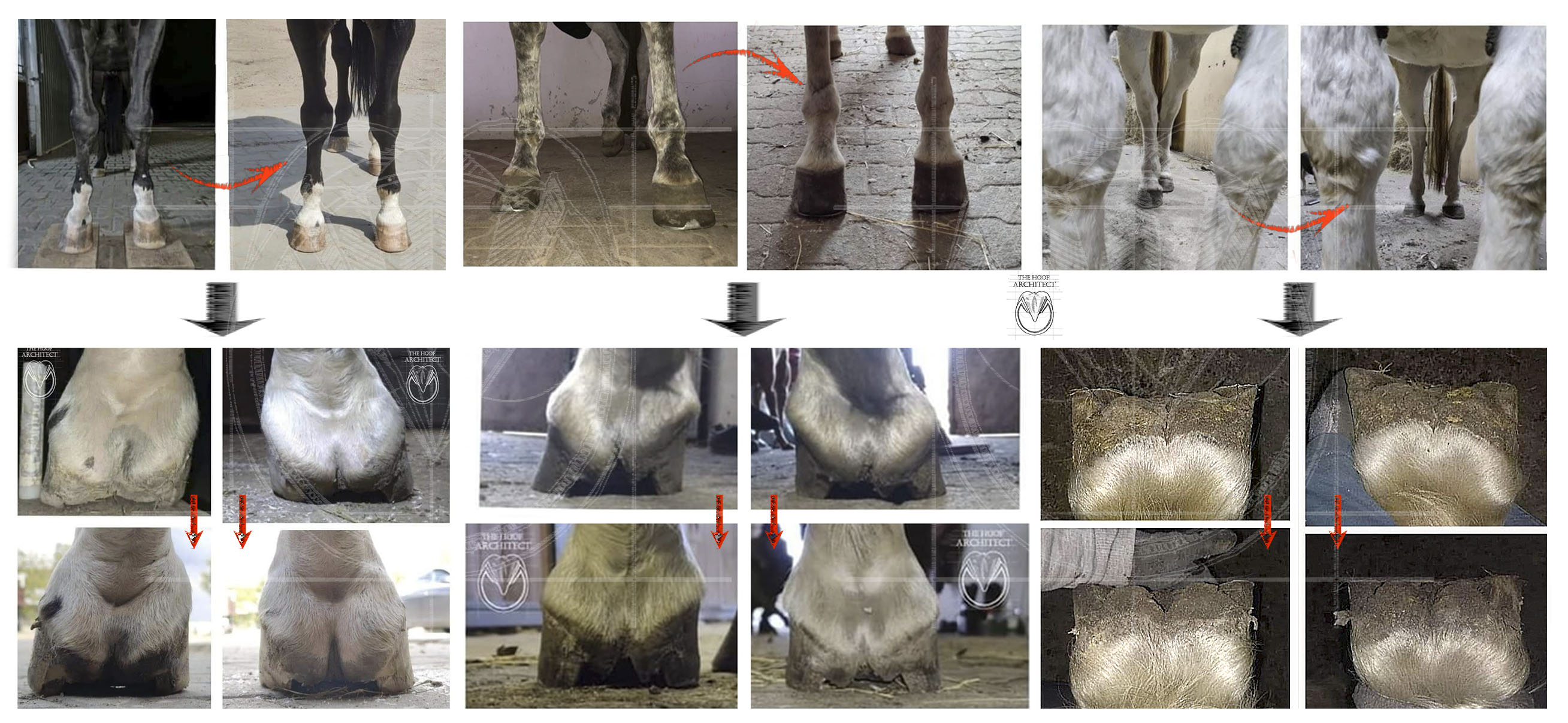
Distal Descent and Its Impact on coronary band shape
Going back to the coronary band distortions. Laminitic horses tend to show very specific distortion patterns which I believe also have to do with the biomechanics of the DIPJ.Distal descent in chronic laminitis multiplies the dorsal and caudal coronary band distortions described previously. When the coffin joint sinks deeper into the horn capsule, the pastern still wants to preserve its range of motion so it pushes down the coronary band and caudal structures out of its way.
The coronary often develops a characteristic dip in the center.
Distal descent and DIPJ extension and flexion
A very common laminitic look of the hoof involves a drop in the central coronary band area. When sinking occurs, P2 is located deeper in the horn capsule. Because range of mediolateral motion in the DIPJ is quite limited, P2 does not interact so much with quarters area. It pushes its way through in the dorsal and caudal aspect of the hoof capsule. |
| Animation showing the pastern pushing the dorsal coronary band down in a case of distal descent. |

A chronic laminitic hoof showing a characteristic drop in the coronary band.
The caudal distortions often resemble those of caudal failure from open heel shoes, but in this case it is sort of a reverse scenario - it is not lack of frog support, the frog is not prolapsed but the walls and cartilages pushed up due to sinking. This is similar to frog prolapse but from the opposite side.
 |
| Animation showing the pastern pushing the caudal structured down in a case of distal descent. |
 |
| A severe chronic laminitic hoof showing the distortion of the caudal hoof related to the distal descent - walls are being pushed up in relation to the internal structures, coronary band vertical and elongated |
What needs to be recognized is that the drop in the coronary band secondary to the distal descent is likely to interfere with the CE measurement. So in an acute case the CE may be big and then seem to decrease, but it is just an illusion. So in chronic laminitis with obvious drop in front, CE may not be a very reliable parameter and the dip itself may be an indicator of previous sinking.
Rotation between the dorsal wall and P3 is also going to exacerbate this drop in the coronary band, as even if the coffin joint preserves its normal range of motion, the whole joint rotated in reference to the dorsal wall and the coronary band, and normal extension on the coffin joint may make the pastern push the coronary band down to a higher extent.

Left: a wide DCA and wide DIPJ extension range hoof with healthy SADP - dorsal wall really thick and strong. Right: a chronic laminitic hoof with coronary band significantly dropped in the center - dorsal wall even thicker.
What about the mild cases?

3 hooves of relatively sound horses, which would struggle with sole depth when barefoot.
- either the quarters being pulled up by the bending of the whole hoof capsule related to large range of flexion of the DIPJ
- or the dorsal area of the coronary band being pushed down by P2 when the DIPJ is in its maximum extension, exaggerated with any degree of distal descent.

Pushed up quarters? Or maybe cranial and caudal hoof is being pushed down with quarters pulled up?
What about limited extension range and distal descent?
Those cases are less common but if that occurs, the coronary groove may become enormously stretched upward when the pastern is not acting over it from above.

2 hooves with limited DIPJ extension range. The coronary groove is elongated due to chronic shearing and force acting directly along the direction of the laminae. No extension of the DIPJ meant no pushing down of the dorsal coronary band.
DDFT tension and determined by that range of DIPJ extension have tremendous influence on what changes are going to occur in the hoof due to laminitis. The influence of DDFT tension on the blood flow within the hoof and its importance in laminitis have been proved by Doctor Redden long ago and should always be taken into account when assessing the severity of the case.DIPJ range of motion and DCA

Left: narrow DCA foot; right: wide DCA foot.
Limb conformation and posture (fetlock joint suspension, DDFT tension) ->
-> DIPJ range of motion ->
-> horn capsule deformation, soft tissue (coronary groove, caudal structures) deformation and displacement, over time also bone remodeling ->
-> horn growing from different oriented coriae ->
-> DCA change
Conclusion: Key Observations
The observations and visual evidence presented in this article highlight a direct correlation between the DIPJ range of motion and the resulting hoof morphology:- DCA and pastern position:
- Limited DIPJ extension effects:
- DIPJ flexion and hoof shape:
- Sole shape and depth:
Summary
The hoof capsule is a viscoelastic record of the forces acting on it over time.The observations presented above suggest that the external hoof capsule and the sole shape and depth are secondary to the functional range of motion within the DIPJ. The DCA serves as a visual indicator of this relationship.
In the following parts the internal anatomy differences between narrow and wide DCA feet will be discussed, to set a foundation for the proposed dorsopalmar hoof types framework introduction.
Bibliography:
Clayton HM, Sha DH, Stick JA, Robinson P. 2007.
3D kinematics of the interphalangeal joints in the forelimb of walking and trotting horses. Veterinary and Comparative Orthopaedics and Traumatology.
Clayton HM. 2010.
Biomechanics of the distal interphalangeal joint. Journal of Equine Veterinary Science, 30(8), 401–405. https://doi.org/10.1016/j.jevs.2010.07.007
Roach JM, Pfau T, Bryars J, Unt V, Channon SB, Weller R. 2014.
Sagittal distal limb kinematics inside the hoof capsule captured using high-speed fluoroscopy in walking and trotting horses. The Veterinary Journal, 202(1), 94–98. https://doi.org/10.1016/j.tvjl.2014.06.014


Comments
Post a Comment