DCA and the Missing Aspects of Dorsopalmar Balance (Part 1 - capsular distortions on macro level)
Unauthorized reproduction, redistribution, adaptation, or derivative works (including redrawn diagrams or similar concepts without attribution) prohibited without written permission.
Introduction
Hoof balance is one of those topics that never seems to lose relevance — and never seems to reach consensus. Ask ten professionals what a “balanced hoof” is, and you will likely hear ten different answers. With modern technology, we now have numerous ways to investigate equine locomotion and hoof biomechanics; yet, many fundamental questions still remain unanswered.
Over the years, numerous systems have been developed to enhance the repeatability of hoof assessment. Specific angles, ratios, mapping systems, reference points, radiographs, and guidelines are used in an attempt to define an optimal hoof. When examined more closely, however, these systems often contradict one another, and in practice, it is frequently impossible to satisfy all of them at the same time. Perhaps this is one reason why many of the most experienced practitioners still rely primarily on their eye and intuition.
Dorsopalmar balance is usually described as a ratio between toe and heel, or as a matter of aligning specific angles and proportions. If these principles were universally valid, certain features should always appear together: tall heels with a steep dorsal wall, low crushed heels with a shallow hoof angle, a horizontal coronary band with a steep hoof angle, and so on. While some hooves do follow these patterns, many clearly do not.
 |
| Fig. 1. Too low or too high heels? Too steep or to low hoof angle? |
The recurring question, then, is not whether these hooves are poorly trimmed, but whether something important is missing from the way we assess them.
One parameter that has received surprisingly little attention is the angle between the dorsal hoof wall and the coronary band in the lateral view — the dorsal hoof wall–coronary band angle (DCA). This angle varies widely between horses, between limbs of the same horse, and over time. It is not directly altered by trimming or shoeing. Instead, it appears to reflect how load is distributed within the hoof capsule, often in relation to posture and distal limb alignment.
In this article, I will focus on DCA as a way to better understand dorsopalmar balance — not as an isolated measurement, but as part of a broader picture that includes hoof capsule distortion, internal load sharing and phalangeal alignment.
Disclaimer: This article is based on long-term clinical observation and experience, not on controlled laboratory studies.
I have already written some posts on DCA in the past. I have since learnt more so in these series I am going to get more into the details.
Part 1.:
https://thehoofarchitect.blogspot.com/2022/07/an-important-hoof-parameter-that-has.html#more
Part 2.:
https://thehoofarchitect.blogspot.com/2022/09/an-important-hoof-parameter-that-has.html#more
What is DCA
 |
| Fig. 2. DCA - Dorsal hoof wall-Coronary band Angle |
DCA is defined as the angle between the dorsal hoof wall and the coronary band when viewed from the lateral aspect. Like many two-dimensional hoof parameters, it is sensitive to perspective, and coronary band distortion can make reference points more difficult to identify. Nonetheless, it is a parameter worth paying attention to.
It is important to understand that DCA can vary by more than 40 degrees between different hooves. It can — and very often does — differ between limbs of the same horse, and it can change over time. Furthermore, it is not directly affected by trimming or shoeing. Changes in DCA appear to occur only as a response to altered load distribution within the hoof, which often follows changes in posture, hoof geometry, or both.
 |
| Fig. 3. DCA value can vary a lot between different feet. |
What Is a 'Correct' DCA?
Before going further, it is worth stating what values I consider appropriate. In the absence of dorsal coronary band distortion (often related to distal descent), DCA values of approximately 100–110° (in the old posts I used to say 100–108°, I have since met quite many good feet around 110°) are typical for front feet I would consider correct or ideal. Hind feet usually fall within a slightly narrower range, around 95–105°.
A hoof with a narrow DCA may remain steep despite removing all heel mass, while a hoof with a wide DCA may retain a low hoof angle despite long or tall heels. This alone causes many systems of assessing hoof balance to break down — especially those that depend on achieving specific toe-to-heel ratios or coronary band–ground angles.
 |
| Fig. 4. Three hooves that fall within narrow, normal and wide DCA range. DCA is one of the four angles of the horn capsule quadrilateral in the lateral view. If this angle can vary so widely and change so substantially, it raises an obvious question: why do we tend to focus on only two of those measurements? |
 |
| Fig. 5. The same hoof before, and after several months. The DCA has changed by 10 degrees. |
Hooves That Do Not Fit the System
 |
Fig. 6. The DCA values of those 2 hooves differ significantly. |
There are hooves that clearly follow conventional expectations of dorsopalmar balance. But there are many that do not.
I consistently observe hooves with long, tall, strong heels and an almost horizontal coronary band, yet with a low hoof angle and a badly broken-back hoof–pastern axis. Conversely, there are hooves with very short, weak, underrun heels and a low coronary band angle paired with a very steep dorsal wall.
In these cases, heel height does not go hand in hand with what we usually call dorsopalmar balance. These feet do not look balanced, regardless of where they fall within measuring or mapping systems. The question then becomes: what is actually being overloaded — the dorsal or the caudal part of the hoof? And what, if anything, should be trimmed or built in height to achieve balance? In many cases, none of the usual options seems valid.
Hoof Shape as a Non-Random Pattern
These hoof shapes are not random. They tend to occur together with specific limb arrangements - particularly phalangeal alignment, pastern length and posture and - but to a lesser degree - hoof management.
Because the horn capsule can deform so extensively, the external appearance of the hoof does not always reveal the internal deformation in an obvious way. It took me months of observation to begin connecting these external shapes with consistent internal distortion patterns.
To make it easier to explain and understand, I have divided the topic into macro and micro level of distortions. All the individual structures within the hoof capsule distort along each other (micro level) creating a overall distortion pattern (macro level) of the whole foot.
 |
| Fig. 7. A simple block will be used to represent a 'normal' hoof morphology. |
The 'Normal' Hoof Is Also a Pattern
Dorsopalmar balance is often considered as a ratio between toe and heel load. Overloaded heels or overloaded toe seem to be the 2 opposite sides of the spectrum, with ‘balance’ laying in the center. But in such a case scenario, where would we put the 2 hooves presented in Fig. 6.?
 |
| Fig. 8. A simplified analogy to how dorsopalmar balance is often viewed: a ratio between the toe and heel load. Where would we place the 2 hooves from Fig. 6.? |
It is worth emphasizing that what we tend to call a “normal” hoof is not necessarily perfectly neutral or distortion-free. It is simply a hoof in which internal load distribution happens to fall within a range that produces proportions we find familiar and visually acceptable.
According to my observations, hooves that fall within the ‘normal’ DCA are usually pleasing to the eye and are easily called balanced. Whilst some of the hooves with DCA on the narrow range may also create a positive impression (sometimes despite some deep internal disfunction!) - hooves with wide DCA are usually the ones most people do not find balanced or healthy, or just feel like there is something off with the whole limb.
A hoof with a DCA in a typical range tends to show:
a hoof–pastern alignment that appears relatively straight,
a balanced relationship between the dorsal wall and the heels both in height and angular relationship,
'healthy looking' heel bulb shape and position.
It is worth mentioning that we do not know whether the 'ideal' hoof morphology is the reason, or a result of the good looking alignment and condition of specific structures. I personally believe that hoof morphology is mostly a mirror of the load distribution coming from above and less the other way round.
Recurring DCA-Related Distortion Patterns
It is very difficult (or impossible) to place hooves with DCA out of range anywhere within the spectrum illustrated in Fig. 8. Because what is overloaded if we have collapsed heels and steep short toe? What is collapsed in a hoof with a long sloping toe and super tall heels?
 |
| Fig. 9. A simplified animation of some of the distortion possibilities on macro level. |
Depending on the DCA range, recurring features may include:
displacement of the caudal aspect of the hoof upward,
displacement of the caudal aspect of the hoof downward,
relative compression of central structures,
or increased loading toward the dorsal or caudal aspects of the hoof, or both at one time.
Many intermediate forms exist, and most hooves display elements of more than one pattern. The important point is that these distortions are organized and repeatable, not random.
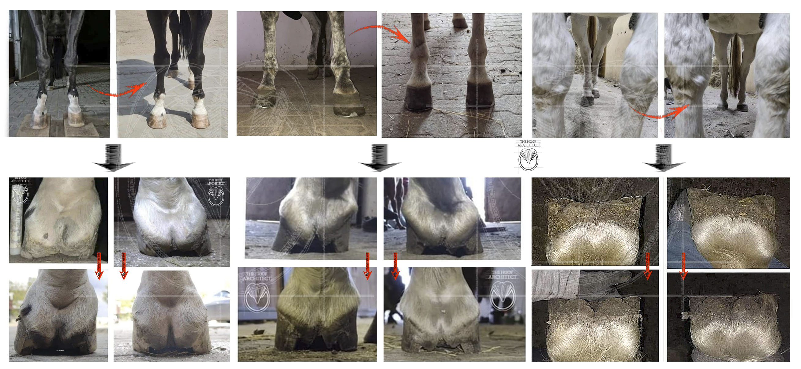
DCA and Longitudinal Hoof Distortion
There is increasing discussion around longitudinal flexion of the hoof when it comes to mediolateral distortions, such as sheared heels. These examples clearly show that the heels can distort independently of what happens at the toe.
 |
| Fig. 10. Each side of the foot can have a different pattern of distortion, thus hooves often present different DCA values on medial and lateral sides. |
However, longitudinal distortion is not limited to mediolateral asymmetry. It is also possible for both heel bulbs to be displaced upward, or both to be pulled downward, producing very different overall hoof geometries despite similar external proportions.
To some degree, DCA allows us to infer which type of internal distortion is present.

Fig. 11. Photomontage of the hoof above. This is a conceptual illustration, not a literal anatomical reconstruction.
On the left - 2 pushed up heels combined in a shape typical of the high DCA hoof.
On the right - in this case, 2 quite normal heel bulbs combined creating a hoof with a normal DCA.
Note the relationship between the pastern width and the distance between the heels - although this is just a photomontage, it is not just a coincidence that it turned out that way. The pastern-hoof capsule relationship is again a crucial factor in determining the overall hoof geometry (more in the following parts).
 |
| Fig. 12. Some of the possible macro distortion patterns with pictures of the corresponding internal structures shapes. |
DCA and Internal Distortion
Dissection pictures with corresponding internal distortion patterns. Once you know what to look at, things start to become more and more apparent. It takes some training to be able to connect the external look of the horn capsule with the internal distortion pattern. DCA is one of the important parameters that help with that.
 |
| Fig. 13. We may learn how to recognize the common internal distortion patterns by assessing the external shape of the hoof capsule. |
More on the internal distortion details in the following parts.
Beyond COP: Load Distribution Within the Hoof
When hoof balance is considered primarily in terms of center-of-pressure (COP) position and movement, a large amount of information may be missed. Load is shared among multiple structures within the hoof capsule, each with different mechanical roles and capacities.
A hoof with COP located centrally may theoretically be:
evenly loaded throughout,
overloaded in the center,
or overloaded cranially and caudally with relative unloading in the center.
 |
Fig. 14. Three hypothetical situations where COP would lie in the middle. |
These are simplified scenarios, but they illustrate that COP alone does not describe how load is actually distributed within the hoof.
Internal Structures and Load Transfer
 |
| Fig. 15. A simplified map of load distribution made based on the observed distortion patterns. |
Looking at distortion patterns led me to think of the hoof as a structure composed of several functional areas.
 |
| Fig. 16. A simplified drawing of the structures transfering the load down over the hoof capsule. |
Load is transferred into and through the hoof capsule by:
the bony column directly,
the deep digital flexor tendon suspending the bony column,
and the caudal structures — including the digital cushion, bars and heels — with collateral cartilages contributing to stabilization.
Depending on the orientation of P2 within the coffin joint, the degree of DDFT tension, and fetlock suspension, load may be transferred more directly onto:
the caudal articular surface of P3 and the navicular bone (P2 upright acting on the caudal aspect of the DIPJ),
the distal border of P3 (P2 acting on the cranial aspect of DIPJ),
or the caudal supporting structures (P2 more horizontal, compressing the structures beneath).
 |
| Fig. 17. A drawing of the main structures sharing the load over the bottom of the hoof capsule. |
 |
| Fig. 18. Structures located in the center of the hoof capsule which may get overloaded in certain scenarios. |
Phalangeal Alignment and Hoof Geometry
 |
| Fig. 19. Hoof-pastern axis and hoof capsule distortion patterns seem to go hand in hand. |
It became increasingly apparent that DCA and three-dimensional hoof capsule distortion patterns strongly correlate with distal limb alignment.
Based on observation of how hooves change shape with altered posture or following injury, my working theory is that phalangeal alignment influences hoof geometry more strongly than hoof geometry influences phalangeal alignment.
There are countless nuanced configurations between “aligned,” “broken forward” and “broken back.” Radiographic examination reveals a wide range of joint relationships that reflect differences in soft-tissue suspension and posture, many of which lie beyond our direct influence. This is going to be covered more extensively in the following parts of the DCA series.
 |
| Fig. 20. A teaser of the second part of these series, which is going to explain how I understand the influence of the fetlock joint biomechanics and DDFT tension on load distribution within the hoof and DCA. I would like to thank the people who have been helping me, inspiring me to keep going with this research and supporting me: Paige Poss, dr Ric Redden, dr Hans Castelijns, members of the EPoP group and others 🙏 |


Comments
Post a Comment