DCA and the missing aspects of the dorsopalmar balance (Part 5: Dorsopalmar hoof types based on limb conformation)
© Ula Krzanowska / The Hoof Architect 2024–2026. All rights reserved.
Unauthorized reproduction, redistribution, adaptation, or derivative works (including redrawn diagrams or similar concepts without attribution) prohibited without written permission.This content, including original observations, biomechanical interpretations, and diagrams, was first publicly presented by the author at the 4th International Congress on Equine Podiatry, Jerez de la Frontera, Spain, May 16–18, 2024.
Unauthorized reproduction, redistribution, adaptation, or derivative works (including redrawn diagrams or similar concepts without attribution) prohibited without written permission.

Introduction to Part 5
In the previous parts of the DCA series (Part 1, Part 2, Part 3, Part 4) we have covered the details of dorsoplmar hoof distortions, fetlock joint and coffin joint biomechanics and internal anatomy related to dorsopalmae balance. In this part we are finally going to connect those aspects together to form a practical framework.
Hoof balance is often viewed in binary terms — as something that is either 'on' or 'off', balanced or unbalanced, as if there were a single, precise threshold beyond which balance is achieved or lost. We frequently talk about 'standard' limbs or limbs with pathology as separate entities requiring entirely different approaches. A 'normal' hoof is treated as one category, while a club foot with DDFT contracture, a limb with dropped fetlock, or any other variation is considered a completely different entity.
Research, biomechanics, and established literature are almost exclusively based on 'normal' horses with 'normal' feet. Descriptions focus on how these 'standard' limbs land, load, and breakover, or how 'standard' feet deform under load. There are specific ‘rules’ established for how certain elements in the limb and the hoof interact with each other to achieve balance.
Research on the biodiversity between individuals depending on conformation is lacking.
Hoof morphology and load distribution within the hoof capsule are commonly described using simplified biomechanical models based on a single, idealised limb configuration. These models typically assume a standard limb geometry and interpret deviations from this norm primarily as pathological variations. However, empirical observation shows substantial diversity in limb conformation, phalangeal alignment, flexor units lengths and stiffness and joint orientation between individual horses. These variations result in measurably different mechanical configurations, which likely influence how forces are transmitted through the digit.
The mechanical equilibrium of the distal limb is governed by the spatial relationship between ground reaction force, skeletal compression, and tensile forces within the flexor tendons and fetlock suspensory apparatus. The moment acting on the distal interphalangeal joint is determined not only by the magnitude of these forces, but also by their moment arms, which are directly influenced by limb geometry, hoof geometry, fetlock position, posture and nuances of phalangeal alignment. Because moment equals force multiplied by moment arm, even small geometric differences may substantially alter the mechanical demand placed on individual structures.
Fetlock joint suspension

Fetlock position plays a central mechanical role in this system. As the fetlock descends, the geometry of the limb changes, altering the relative position of the ground reaction force and the mechanical demand placed on the deep digital flexor tendon. Depending on the specific configuration, fetlock descent may increase elongation-driven tension in the DDFT due to suspension of body weight, while simultaneously reducing the ground reaction force moment arm relative to the distal interphalangeal joint by shifting the center of pressure caudally. These effects occur simultaneously, and their relative magnitude depends on limb conformation, tendon properties, hoof geometry and joint orientation.
An element I have never seen mentioned, is that fetlock suspension affects how the weight is being distributed within the hoof capsule - whether it is going to be concentrated in the middle (under the boney column in case of upright pasterns), distributed more evenly or spread between the toe and heels.
When the fetlock is upright, more load is being transferred directly down through the bones, limiting the role of tendons and ligaments in suspending the weight. The lower it drops, the more vertical load is transferred into partially horizontally suspended load. It also changes the gait quality and amount of shock absorption.
Therefore, there is no specific direction in which certain fetlock positions are going to shift COP towards, but it determines how spread or how concentrated that load is going to be - which has tremendous influence on hoof morphology.
DDFT tension
 Load distribution depending on DDFT tension. Left: low DDFT tension, load concentrated in the caudal area of the foot. Middle: neutral DDFT tension, load spread in the middle. Right: High DDFT tension, load concentrated at the toe. The thickness of the red lines reflects the tension, not the real thickness of the structure.
Tension within the DDFT may arise from different mechanisms. Contraction-driven tension originates from active muscle contraction and serves primarily to stabilise the phalangeal column. Elongation-driven tension results from passive stretching of the tendon due to external loading and suspension of mass. In addition, congenital or early-acquired shortening of the muscle–tendon unit alters the baseline length–tension relationship and may produce persistent tension independent of current neuromuscular conditions. These distinct mechanisms may produce superficially similar external appearances while reflecting fundamentally different internal mechanics.
The position of the fetlock joint alters the mechanical demand placed on the DDFT by changing joint geometry and moment arms. This effect occurs regardless of whether the underlying cause is active muscle contraction, passive loading, or structural shortening of the tendon unit. As a result, fetlock position does not merely reflect tendon tension but also participates in determining how forces are distributed within the digit.
DDFT tension and COP - what is the cause and what is the result?
DDFT tension creates a flexor moment at the distal interphalangeal joint (DIPJ), which is traditionally described to be balancing the extensor moment generated by the ground reaction force (GRF) acting around the center of pressure (COP), located cranial to the center of rotation (COR) of the DIPJ. This is the conventional biomechanical explanation of distal limb equilibrium in the equine hoof.
 |
| Grey arrow shows the GRF acting on the normal hoof when the hoof is stationary. Drawing by Andrew Parks, source: https://www.equipodiatry.com/news/articles/aspectsoffunctionalanatomyofthedistallimbhtml |
There is research showing that hoof growth often leads to lowering of the palmar angle (PA), breaking back of the hoof–pastern axis (HPA), and increasing toe length relative to the COR, all of which increase DDFT demand. Mathematical calculations have been performed to try and describe how manipulating hoof geometry influences the deep flexor demand.
The problem arises when these findings are extrapolated to the general horse population as absolute rules, as if a single universal baseline existed to which all horses could be compared. Most of these studies measure RELATIVE changes from an individual baseline established within a specific animal. There is no universal baseline set of the whole population of horses to start calibrate the measurements. DDFT tension is a result of the baseline tension AND ON TOP OF THAT - NOT INSTEAD OF THAT - the hoof geometry.
It is widely understood that increasing PA reduces the flexor moment demant at the DIPJ relative to the starting point, but that starting point is rarely defined.
The COP - DDFT tension relationship goes both ways, but it is rarely considered how the baseline DDFT tension/length sets the starting point for each individual horse.
What exactly does this mean in practice?
Let us imagine two feet with different initial configurations: a club foot with a hyperpositive PA and broken-forward HPA, and a bull-nosed negative palmar angle (NPA) foot with a severely negative PA and broken-back HPA.

If both feet are trimmed or shod to achieve the same PA, the same HPA alignment (at least aligning P3 and P1), and similar proportions around the COR:
- Can we assume that these two limbs will now have the same load distribution between the DDFT, superficial digital flexor tendon (SDFT), and suspensory ligament (SL)?
- Can we assume they are now biomechanically equivalent, with the same COP position?
- Do we expect them to grow evenly, or at least to grow in the same way over time?
- Do we expect their biomechanics to be the same simply because their geometry was made similar?
My interpretation of their resulting load distribution and subsequent growth tendencies is as follows:


 |
| Alignment of the digit is a moment in time. 2 similarly aligned after the trim hooves can have very different load distribution within, very different COP locations, different biomechanics, problems, tendencies and different needs. |
Recognizing that each horse has an individual baseline level of DDFT tension, which influences limb posture and hoof mechanics, has been an important component of the work of Doctor Ric Redden.

Drawings by Dr Redden. Dr Redden has been teaching about the role of DDFT tension in shifting the load cranially or caudally for decades.
My observations suggest that baseline DDFT tension has very substantial influence on COP location, more than base of support ratio or any other parameter.
Fetlock position and DIPJ moment equilibrium
However, as said before, DDFT tension and fetlock position influence each other. Descending fetlock shifts the weight caudally (COP moves caudally) while simultaneously placing more load on the DDFT due to leverage increase (COP moved cranially). Depending on the conformational nuances and baseline DDFT tension, caudal or cranial shift of habitual COP location is possible as a result.
COP travels along the solar surface during different phases of the stance phase. After landing it is located in the caudal area and shifts cranially as the body of the horse travels forward above the limb. Different conformational configurations may alter how long the COP stays behind, under and in front of COR during the stance phase.
As long as COP is located behind COR, GRF does not exert an extensor moment on the DIPJ, but in fact it acts a flexor moment. When the DDFT tension is insufficient to shift COP forward early enough, the GRF contributes to the flexor moment, reducing the mechanical demand on the DDFT as the primary flexor moment generator.
Insufficient DDFT tension to counterbalance the fetlock descent: In cases of DDFT rupture or laxity, the DDFT tension may not be sufficient enough to counterbalance the extensor moment exerted by the extensor branches of the SL and descending fetlock. When toe lifts off the ground, it indicates that COP location is behind COR, thus GRF acts as a flexor, not extensor moment during standing. It may change with the limb placement position - the toe may float when the limb is placed more forward but not when it's moved back. These are the very severe cases, but it seems fair to assume that all horses fall within the spectrum between COP location being too far forward and too far backward, with the 'ideal' ones in the center of the spectrum.
 |
| A total collapse of the fetlock suspension and DDFT tension. Photo courtesy of Dr Raul Bras. |
 |
| Lever of the pastern is one of the factors contributing to the toe raising off the ground when the DDFT tension is insufficient |
.

Caudal structures act as a leverage fulcrum for the descending pastern and get compressed, weight shifts onto the palmar processes of P3. If the last point of ground contact is not far back enough in reference to COR, the arm of the flexor moment may not be sufficient to keep the toe on the ground (extending the base of support extends that moment arm and helps in those cases).
In the opposite case scenario, where the DDFT tension is higher than ‘normal’, COP shifts further cranially and does not allow the caudal structures to be loaded properly, concentrating all the weight on the toe and bringing the heels up in the air, no matter the base of support ratio.


On the left: DDFT tension insufficient. On the right: descending fetlock loading the DDFT to the point of heels rising.
Looking at the end-of-spectrum cases
If we believe all feet and limb fall somewhere within the DDFT tension/fetlock suspension spectrum, it seems valuable to look deeper into the end-of-spectrum cases, to see the exaggerated result of those factors being away from the neutral (middle of spectrum) state. Once we understand what to look for, it becomes easier to spot the subtle cases too.
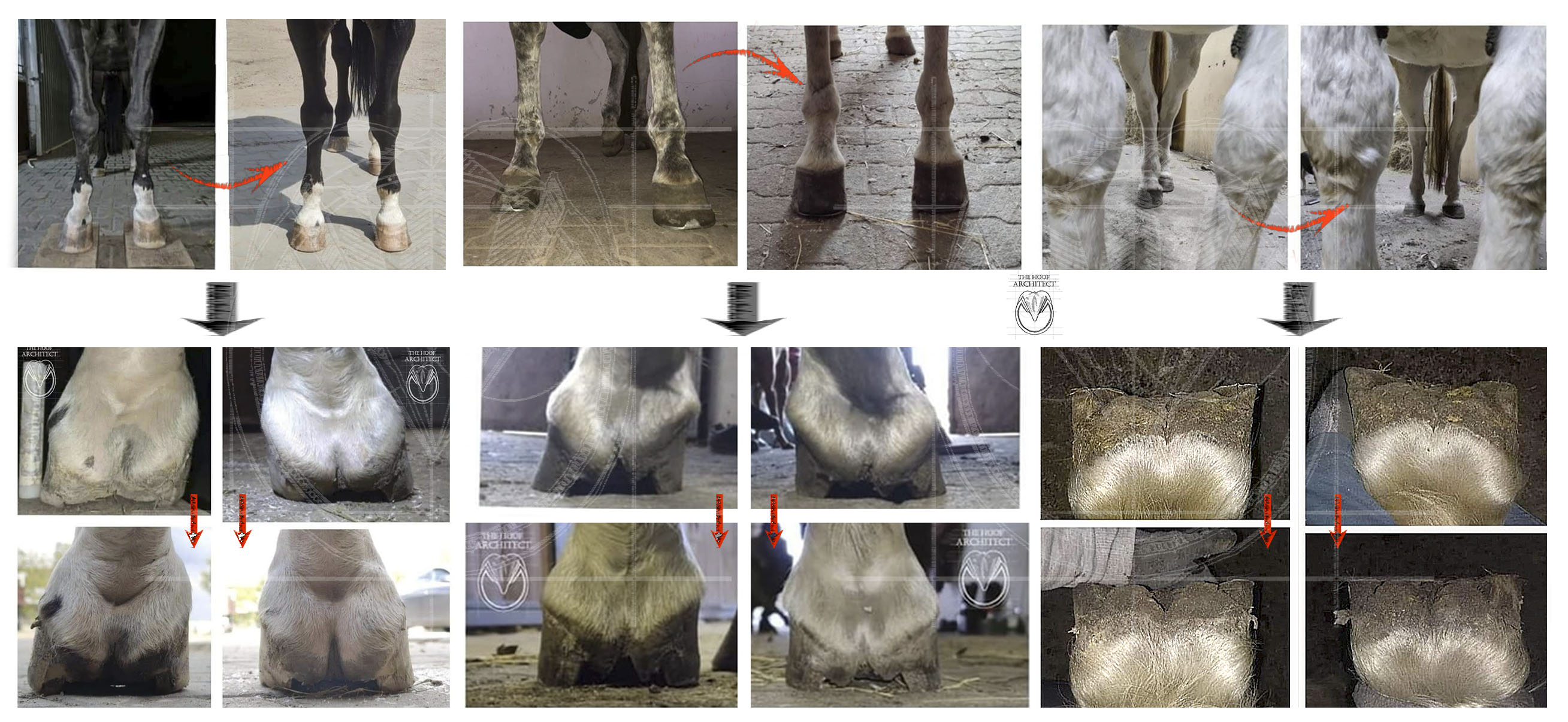
Looking at flexural deformities in foals may give us a hint what configurations may be possible. Are all of those flexural deformities resolved before the horse matures? Are they resolved fully, or does a subtle blueprint of the early conformation stay there in some of the cases?


Hoof capsule morphology primarily reflects the long-term mechanical state of the system. Because the hoof capsule is a viscoelastic structure, repeated loading within specific mechanical configurations results in time-dependent deformation and growth adaptation, according to the habitual position of the center of pressure and pressure distribution. Morphological features such as heel collapse, central hoof collapse, dorsal wall curvature, or changes in DCA may therefore represent the cumulative mechanical history of the limb rather than isolated pathological events.
Considering the amount of congenital or early acquired developmental conformational traits along with possible injuries, degeneration and compensation patterns, we may observe huge diversity not only between hoof shapes, but also limb conformations and biomechanical configurations.
The so-called ‘normal’ feet fall somewhere in the middle of the spectrum of possible fetlock and DDFT tension configurations.
Let’s compare some of the end-of-spectrum cases in adult horses.

Why propose a new framework
Current biomechanical research has largely focused on limbs considered mechanically normal, and there is limited experimental data directly comparing force distribution, center of pressure location, and tendon loading between limbs with different conformational configurations, such as club feet, limbs with chronically dropped fetlocks, or limbs with structural tendon shortening. As a result, many existing mechanical models are derived from a limited subset of limb configurations and may not fully capture the mechanical diversity observed in the equine population.
The interpretation presented here represents a biomechanical framework based on established mechanical principles, including moment equilibrium, force transmission, and viscoelastic deformation, combined with empirical observation of hoof morphology and limb conformation. Within this framework, hoof morphology can be understood as a mechanical record of how forces are resolved within the limb over time.
This approach does not assume a single ideal limb configuration as a universal goal, but instead recognises that each limb operates within its own mechanically defined equilibrium state. Considering the combinations of DDFT tension and fetlock position, this framework matrix produces nine (3x3) configurations that describe possible tendencies and directions of hoof distortion in response to load distribution.
DDFT tension - fetlock suspension relationship and DIPJ biomechanics
These 2 aspects play together, determining the ultimate outcome. Their relationship is very important when it comes to the alignment and range of motion in the DIPJ, which is a factor directly correlated to the DCA value, to the COP location trajectory throughout the stance phase and when standing.
- The more DDFT tension and the lower the fetlock, the more flexion-dominant the DIPJ biomechanics
- The less DDFT tension and the more upright the fetlock, the more extension-dominant the DIPJ biomechanics
The 3×3 matrix of biomechanical configurations
Rather than existing as a single mechanical norm, limb configurations form a continuous spectrum of equilibrium states. These states arise from variation in tendon length and stiffness, joint geometry, phalangeal alignment, and fetlock position. So-called 'normal' feet represent positions within this spectrum, not a universal reference configuration. At one extreme, limbs with structurally shortened flexor units exhibit increased baseline tendon tension and characteristic capsule morphology. At the opposite extreme, limbs with increased fetlock descent and reduced baseline tendon tension exhibit different but equally predictable morphological adaptations.

Between these extremes exists a continuum of intermediate configurations. The hoof capsule adapts accordingly.
A shift in interpretation
This framework does not define a single ideal hoof form. Rather, it recognizes that different conformations produce different equilibria. Hoof morphology should first be interpreted properly, to then decide whether it needs correction, and if yes - why and how. DCA provides a window into that mechanical reality but not as an isolated parameter. It serves as part of a mechanically coherent system linking limb conformation, force distribution, and capsule adaptation.
Numerous other factors that influence hoof morphology
When discussing factors influencing hoof morphology, focus is often placed on local or external features of the hoof capsule. This represents a symptomatic approach, addressing the visible manifestation rather than the underlying mechanical cause. Numerous factors influence hoof morphology: genetics, developmental influences, SADP quality, horn thickness and stiffness, soft tissue properties, movement quantity and quality, and substrate characteristics. Hoof care also has a substantial influence — trimming, shoeing, surface of support, choice of materials - these all affect how forces are transmitted.
It is very uncommon for a hoof to transition from one distortion pattern tendency to another without a significant change in the biomechanical equilibrium of the limb:
- A flat bull-nosed foot will not become a club foot unless the deep digital flexor unit contracts.
- A club foot will not become a low-angled foot unless there is rupture, elongation, or surgical alteration of the deep digital flexor unit.
- An elongated (wide DCA) foot will not become short and compressed (narrow DCA), unless the fetlock drops down or DIPJ extension range becomes limited due to change of posture, degeneration of injury.
Two feet may present identical angulation and phalangeal alignment immediately after trimming, yet grow in completely different ways over the cycle. Distortion patterns become most evident in feet that went for some time without correction and that had not been corrected according to their needs.
Conclusion
This framework is meant to help recognize different and repeatable patterns of hoof distortions, in order to understand what problems we may be dealing with, what forces are we fighting, where the potential pathologies may be expected, what intervention may be needed and what limitations to expect. It may help predict growth tendencies, localize areas that need special protection and help develop a more comprehensive plan to help each individual limb in the best possible way.
In the next part the 9 types will be described, covering the aspects mentioned above.


Comments
Post a Comment